Automated Mineralogy Laboratory
The Automated Mineralogy Laboratory is a research laboratory within the Center for Subsurface Earth Resources and the Department of Geology and Geological Engineering that is dedicated to mineral characterization and application development for the minerals, energy, environmental, biological, and planetary research groups and industries. The laboratory is equipped with a TESCAN Integrated Mineral Analyzer (TIMA), a revolutionary instrument used for minerals and materials analysis.
Automated mineralogy utilizes Scanning Electron Microscopy (SEM) and Energy-dispersive X-ray spectroscopy (EDS) to perform rapid and accurate analysis of mineralogical and industrial samples for research and industry. The focus of the laboratory is to provide improved understanding of materials in order to better predict their management, development, and the effective recovery of resources.

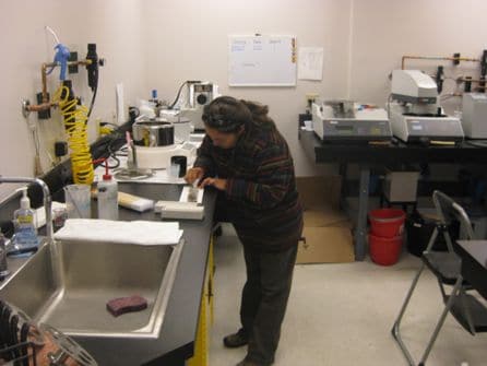
The Automated Mineralogy Facility is also equipped with a fully operational sample preparation facility. Automated state of the art equipment ensures the efficient and safe preparation of a variety of materials, from dust-sized particles to rock samples. The sample preparation facility has both, water- and alcohol-based polishing units, allowing us to work with water-soluble compounds.
Contact
Department of Geology and Geological Engineering
Colorado School of Mines
1516 Illinois Street
Golden, CO 80401
Dr. Katharina Pfaff
kpfaff@mines.edu
303-384-2487
Fax: 303-273-3859
Applications
Automated Mineralogy provides quantitative mineralogical and textural data, false-color mineral maps, and robust statistical data than can be used to quantify a variety of important variables.
- Highly accurate mineral (phase) abundance (i.e. modal abundance) maps
- Modal Mineral/Phase Abundance Data (in Mass% and Area%)
- Particle, Grain and Pore Size distribution
- Element Distribution Maps
- Elemental Deportment
- Element Mass
- Mineral Association
- Porosity/Organic matter quantification
- Mineral/Phase locking and liberation characteristics
- Lithotyping
Mineral Exploration and Mining
Quantitative mineral analysis has a wide variety of applications in the mining and minerals sector. At the Automated Mineralogy Facility at the Colorado School of Mines, research is focused on mineral identification and quantification for the recovery of base metals, precious metals and critical minerals.


Energy and Petroleum Resources
The Automated Mineralogy Facility at the Colorado School of Mines provides important solutions for Oil and Gas reservoir characterization. Mineralogy, porosity and organic matter measurements can be carried out for a variety of rock types including sandstone, fine-grained oil shale, carbonate, coal and more.


Heavy Mineral Analysis
Automated mineralogy is a powerful tool for many other areas including astrobiology, palaeoclimate reconstruction, provenance analysis and mineral indicator studies.

Natural and Synthetic Materials, Quality Control, Environment and Biology, Metallurgy and Engineering
Instrumentation

The TESCAN Integrated Mineral Analyzer (TIMA) is a fully automated SEM-based analysis system that provides quantitative mineralogical and textural data on the basis of automated point counting. The instrument contains a custom-built electron-beam platform equipped with four energy dispersive X-ray spectrometers (EDS) for mineral and compound identification within a wide range of sample types.
The TIMA software allows for the automated stepping of the electron beam across samples at a user-defined pixel resolution (typically 1 – 40 micrometers). At each pixel, the system collects a backscatter electron (BSE) signal and an EDS spectrum. A mineral or phase identification is made on the basis of the BSE value and elemental intensities.
These include:
- Highly accurate mineral (phase) abundance (i.e. modal abundance) maps
- Modal Mineral/Phase Abundance Data (in Mass% and Area%)
- Particle, Grain and Pore Size distribution
- Element Distribution Maps
- Elemental Deportment
- Element Mass
- Mineral Association
- Porosity/Organic matter quantification
- Mineral/Phase locking and liberation characteristics
- Lithotyping
The TESCAN Integrated Mineral Analyzer is designed to accommodate
- standard thin sections (27 x 46 mm)
- 30mm and 25mm epoxy mounts
Samples are analyzed using automated scanning electron microscopy (automated mineralogy) at the Colorado School of Mines. Sample are loaded into the TESCAN-VEGA-3 Model LMU VP-SEM platform and analysis is initiated using the control program TIMA3.
Four energy dispersive X-ray (EDX) spectrometers acquire spectra from each point or particle with a user defined beam stepping interval (i.e., spacing between acquisition points), an acceleration voltage of 24 keV, and a beam intensity of 14. Interactions between the beam and the sample are modeled through Monte Carlo simulation. The EDX spectra are compared with spectra held in a look-up table allowing a mineral or phase assignment to be made at each acquisition point. The assignment makes no distinction between mineral species and amorphous grains of similar composition. Results are output by the TIMA software as a spreadsheet giving the area percent of each composition in the look-up table. This procedure allows a compositional map to be generated. Composition assignments will be grouped appropriately.
Scan Types
The TESCAN Integrated Mineral Analyzer (TIMA) can perform several different scan types to best suit the needs of each client. Each of these scan types is listed below, and includes a brief description of the primary uses for each type.
- Modal Analysis: Modal analysis is designed for detailed evaluation of minerals and their proportions in the sample. Pixel-by-pixel processing allows easy detection of very fine variations in the mineral or chemical composition and provides information necessary for precise analysis of assayed samples. Produces full false-color images of sample/thin section.

- Liberation Analysis: Liberation analysis is dedicated to analysis of particles and grains in the sample and provides information about their size, density, texture, as well as mineral and elemental composition. Grain-by-grain processing of all particles in assayed samples allows detection and evaluation of various types of ore particles and offers data essential for i.e., ore processing control or heavy mineral studies.

- Bright Phase Search: Bright Phase Search is focused on the fast search for a specific mineral or group of particles of interest (i.e., datable minerals, precious metals or rare earths, usually forming a small percentage of the total number of particles in the sample). For these minerals or particles, it provides information about size, density, texture, as well as mineral and elemental composition, which can be further used for process optimization and ore processing efficiency control.


TIMA Specifications
TESCAN Integrated Mineral Analyzer (TIMA) is based on a VEGA thermal emission scanning electron microscope. Special VEGA column design with permanent gun high-vacuum and the isolation valve significantly increases emission stability and tungsten filament lifetime.
A computer controlled stage carries a specially designed mineralogical sample holder. It allows inserting up to 7 resin blocks with a diameter of 30 mm or 25 mm at the same time or up to two standard petrographic thin sections.
In the center of the holder, an EDX/BSE calibration standard for automatic system calibration is placed. The standard consists of a platinum Faraday cup for BSE signal calibration and manganese standard for system performance checks.
Technical Specifications
The TESCAN Integrated Mineral Analyzer (TIMA) is equipped with an ultra-fast YAG scintillator BSE detector, which is one of the key components of the data acquisition part of the system.
The BSE detector is complemented with four silicon drift EDX detectors (PulseTor) in analytical geometry to cover maximum solid angle of X-ray data acquisition for high throughput analysis.
The SE detector is a Everhart-Thornley detector.
Sample Preparation

In addition to the instrumentation laboratory, the Automated Mineralogy Facility at the Colorado School of Mines is equipped with a fully operational sample preparation facility. Automated state of the art equipment ensures the efficient and safe preparation of a variety of materials, from dust-sized particles to 10-cm-sized rocks. The Automated Mineralogy Facility has both water- and alcohol-based polishing units, allowing us to work with water-soluble compounds as well.
The following sample types can be prepared in our sample preparation facility:
- Particulate material: These include mill products, sediment samples, and heavy mineral separates
- Whole-rock material: Samples of nearly any size (max dimensions: 7.5 x 10 x 2 cm) can be cut and polished, or mounted in epoxy
- Compounds and alloys: These include industrial materials and metallurgical compounds
- Carbon tape mounts: Used for mounting coarse-grained material that does not require grinding or polishing prior to analysis
Quality assurance and quality control are the top priorities in sample preparation. From cutting or splitting to mounting, polishing, and coating, high-quality samples are produced during all stages of preparation.
Dry Laboratory Equipment
- Ductless dust hood for fine-grained sample preparation
- Rotary Micro Riffler for splitting samples into representative aliquots
- Hydrosonic bath for sample disaggregation
- Large oven for sample drying
- Qake machine for rapid and thorough sample mixing
- Electronic balance for precise sample measurement
- Carbon coater for establishing electrical conductivity of each sample
- Sized fractions of graphite for sample mounting
Wet Equipment
- Two Struers Tegra grinding and polishing stations (water and alcohol based) with diamond polishing grits
- Struers precision diamond wheel cut-off saw and grinding machine for specimen preparation
- Optical microscope and camera to ensure sample quality
- Molds, resins and hardeners for a variety of sample shapes and sizes
Pricing
The Qemscan Facility uses fixed hourly pricing for scan time on the instrument. Per-hour scanning charges are based on a two-tier structure. Other services, such as sample preparation, are charged at cost.
| Sample Preparation | Price |
|---|---|
| Epoxy mounts (30 mm) | $50 per sample |
| Polished rock slabs | $150 per sample |
| Polished epoxy blocks | $150 per sample |
| Carbon coating | $5 per coating cycle |
| Scanning Charges | Price |
| Research rate* | $75/hr beam time |
| Commercial rate | $200/hr beam time |
| Other Rates | Price |
| Data reduction and reporting | $75/hr |
| Development work** | $75/hr |
| Facilities and usage fee*** | 8.5% of invoice sub-total |
*The research rate only applies to academic research projects supported by Mines and accredited universities. Please contact the laboratory to see if your project qualifies for the research rate.
**Development work applies to any time spent in the development of new mineral definitions, new sample preparation techniques, and any new methods of scanning not currently employed in the Qemscan Facility.
***This fee is charged to all non-Mines projects. Research projects endorsed and sponsored by Mines are exempt from this fee.
Publications
Selected publications based on data acquired at the Automated Mineralogy Facility (TIMA and QEMSCAN) at the Colorado School of Mines:
- Nicco, M., Holley, E.A., Hartlieb, P., Kaunda, R., Nelson, P.P., 2018, Methods for Characterizing Cracks Induced in Rock. Rock Mechanics and Rock Engineering, 51, 2075-2093.
- Qiu, K-F., Marsh, E., Yu, H-C., Pfaff, K., Gulbransen, C., Gou, Z-Y., Li, N., 2017, Fluid and metal sources of the Wenquan porphyry molybdenum deposit, Western Qinling, NW China. Ore Geology Reviews, 86, 459-473.
- Sliwinski, J.T., Bachmann, O., Dungan, M.A., Huber, C., Deering, C.D., Lipman, P.W., Martin, L.H.J., Liebske, C., 2017, Rapid pre-eruptive thermal rejuvenation in a large silicic magma body: the case of the Masonic Park Tuff, Southern Rocky Mountain volcanic field, CO, USA. Contributions to Mineralogy and Petrology, 172, 30.
- Peng, W., Wang, Z., Song, Y., Pfaff, K., Luo, Z., Nie, J., Chen, W., 2016, A comparison of heavy mineral assemblage between the loess and the Red Clay sequences on the Chinese Loess Plateau. Aeolian Research, 21, 87-91.
- Hendrickson, M.D., Hitzman, M.W., Wood, D., Humphrey, J.D., Wendlandt, R.F., 2015, Geology of the Fishtie deposit, Central Province, Zambia: iron oxide and copper mineralization in Nguba Group metasedimentary rocks. Mineralium Deposita, 50, 717-737.
- Capistrant, P.L., Hitzman, M.W., Wood, D., Kelly, N.M., Williams, G., Zimba, M., Kuiper, Y., Jack, D., Stein, H., 2015, Geology of the Enterprise Hydrothermal Nickel Deposit, North-Western Province, Zambia. Economic Geology, 110, 9-38.
- Kuiper, Y.D., Williams, P.F., Hmilton, M.A., 2015, Effects of Paleogene faults on the reconstruction of the metamorphic history of the northwestern Thor-Odin culmination of the Monashee complex, southeastern British Columbia. Lithosphere, 7, 321-335.
- Lynch, K.L., Horgan, B.H., Munakata-Marr, J., Hanley, J., Schneider, R.J., Rey, K.A., Spear, J.R., Jackson, W.A., Ritter, S.M., 2015, Near-infrared spectroscopy of lacustrine sediments in the Great Salt Lake Desert: An analog study for Martian paleolake basins. Journal Geophysical Research: Planets, 599-623.
- Wang, X., Geysen, D., Padilla Tinoco S.V., D’Hoker, N., Van Gerven, T., Blanpain, B., 2015, Characterisation of copper slag in view of metal recovery. Mineral Processing and Extractive Metallurgy, 124, 65-124.
- Nie, J., Peng, W., 2014, Automated SEM-EDS heavy mineral analysis reveals no provenance shift between glacial loess and interglacial paleosol on the Chinese Loess Plateau. Aeolian Research, 13, 71-75.
- Wunsch, A., Navarre-Stichler, A.K., Moore, J., Ricko, A., McCray, J.E., 2013, Metal release from dolomites at high partial-pressure of CO2. Applied Geochemistry, in press.
- Nie, J., Peng, W., Pfaff, K.,Möller, A. Garzanti, E., Andò, S., Stevens, T., Bird, A., Chang, H., Song, Y., Liu, S., Ji, J., 2013, Controlling factors on heavy mineral assemblages in Chinese loess and Red Clay. Palaeography, Plaeoclimatology, Palaeoecology, 381-382, 110-118.
- Sharma, R., Prasad, M., Batzle, M., Vega, S., 2013, Sensitivity of flow and elastic properties to fabric heterogeneity in carbonates. Geophysical Prospecting, 61, 270-286.
- Chin, K., Pearson, D., Ekdale, A.A., 2013, Fossil Worm Burrows Reveal Very Early Terrestrial Animal Actvity and Shed Light on Trophic Resources after the End-Cretaceous Mass Extinction. Plos One, 8, 1-8.
- Gregory, M.J., Lang, J.R., Gilbert, S., Hoal, K.O., 2013, Geometallurgy of the Pebble Porphyry Copper-Gold-Molybdenum Deposit, Iliamna, Alaska. Economic Geology, 108, 483-494.
- Lesher, E.K., Honeyman, B.D., Ranville, J.F., 2013, Detection and characterization of uranium-humic complexes during 1D transport studies. Geochimica et Cosmochimica Acta, 109, 127-142.
- Schmandt, D., Broughton, D., Hitzman, M.W., Plink-Bjorklund, P., Edwards, D., Humphrey, J., 2013, Economic Geology, 108, 1301-1324.
- Tinnacher, R.M., Nico, P.S., Davis, J.A., Honeyman, B.D., 2013, Effects of Fulvic Acid on Uranium (VI) Sorption Kinetics. Environmental Science & Technology, 47, 6214-6222.
- Zhao, X.-F., Zhou, M.-F., Hitzman, M.W., Li, J.-W, Bennett, M., Meighan, C., Anderson, E., 2012, Late Paleoproterozoic to Early Mesoproterozoic Tangdan Sedimentary Rock-Hosted Strata-bound Copper Deposit, Yunnan Province, Southwest China. Economic Geology, 107, 357-375.
- Wandler, A.V., Davis, T.L., Singh, P.K., 2012, An Experimental and Modelig Study on the Response to Varying Pore Pressure and Reservoir Fluids in the Morrow A Sandstone. International Journal of Geophysics, Article ID 726408, 17 pages.
- Mahan, K.H., Schulte-Pelkum, V., Blackburn, T.J., Bowring, S.A., Dudas, F.O., 2012, Seismic structure and lithospheric rheology from deep crustal xenoliths, central Montana, USA. Geochemistry, Geophysics, Geossystems, 13, 1-11.
- Barnhart, K.R., Mahan, K., Blackburn, T.J., Bowring, S.A., Dudas, F.O., 2012, Deep crustal xenoliths from central Montana, USA: Implications for the timing and mechanisms of high-velocity lower crust formation. Geosphere, 8, 1408-1428.
- Ault, A.K., Flowers, R.M., Mahan, K.H., 2012, Quartz shielding of sub-10 um zircons from radiation damage-enhanced Pb loss: An example from a metamorphosed mafic dike, northwestern Wyoming craton. Earth and Planetary Science Letters, 339-340, 57-66.